Brain Tumours
Brain Tumours are either primary or secondary in type.
Primary Brain Tumours grow from the brain itself, or from its encasing membranes. Some examples of some of the names used for primary brain tumours include glioma, astrocytoma, glioblastoma, oligodendroglioma, ependymoma, pituitary adenoma, meningioma. This list includes some of the more common ones, although there are many others not listed here.
Secondary Brain Tumours are tumours that have spread to the brain via the blood stream, such as from the lung, breast, skin or bowel.
The treatment of Brain Tumours is different in every case. Some of the factors that Gavin Davis takes into account in determining the most appropriate treatment in each patient include:
- Tumour type
- Tumour size
- Tumour location (which part of the brain is involved)
- Multiplicity - Whether there is one or more than one tumour
- Patient age
- General health of the patient, and the presence of other diseases/illnesses
- Neurological status of the patient (is the patient alert and talkative, or is the patient deeply unconscious or hemiplegic)
- Patient’s wishes
- Previous treatment, including radiotherapy
Once all of these factors have been analysed, a treatment plan is developed for the patient. This treatment plan may include:
Furthermore, if you have had seizures, your management will also include the use of anticonvulsant medications (whether or not you have had surgery).
It should be noted in all patients diagnosed with a brain tumour, that they are not allowed to drive a motor vehicle or operate heavy machinery, and must notify the local driver's licence authority regarding this. Once treatment has been completed, and after consultation with your doctor, a determination will be made regarding future capacity to drive.
- Surgical Resection - surgery to remove the tumour via a craniotomy (see illustration below)
- Surgical Biopsy - surgery to remove a small sample of the tumour
- Radiotherapy or radiosurgery - radiation used to shrink or slow tumour growth
- Chemotherapy - drug treatment used to shrink or slow tumour growth
Furthermore, if you have had seizures, your management will also include the use of anticonvulsant medications (whether or not you have had surgery).
It should be noted in all patients diagnosed with a brain tumour, that they are not allowed to drive a motor vehicle or operate heavy machinery, and must notify the local driver's licence authority regarding this. Once treatment has been completed, and after consultation with your doctor, a determination will be made regarding future capacity to drive.
Each patient may require one or more of these treatments. For example, some tumours need surgery and radiotherapy and chemotherapy. Others may need only surgery or only radiosurgery.
Your treatment will be determined after Prof Davis has reviewed all the relevant information, and has consulted with a number of other professionals, expert in the management of brain tumours. At Cabrini Hospital, Prof Davis utilises the services of the multidisciplinary brain tumour group to ensure that you receive the most appropriate, current, reliable treatment for your tumour. Some of those involved in the management include a neuro-oncologist, brain tumour nurse, radiation oncologist, rehabilitation physician and pathologist. Depending upon the pathological type of tumour, some tumours can be cured by surgery alone (for example, many meningiomas), whilst other tumours can only be controlled with surgery and other treatments (for example, glioblastoma). Once your pathology results are available, Prof Davis will discuss the prognosis and management strategies with you.
For further information on Clinical Guidelines, click here
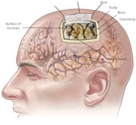
This image demonstrates a craniotomy, or opening of the skull, to expose the meninges (membranes) and the underlying brain and blood vessels. The site of the craniotomy on the head is modified in each case, based on the CT and MRI findings.
This illustration reproduced with permission from the Craniotomy pamphlet (RACS, NSA, Mi-tec Medical Publishing). The complete pamphlet is available from your surgeon.
This illustration reproduced with permission from the Craniotomy pamphlet (RACS, NSA, Mi-tec Medical Publishing). The complete pamphlet is available from your surgeon.
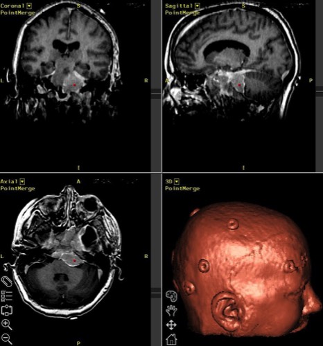
The image above demonstrates a tumour displayed with the stereotactic 3D reconstruction of the head.
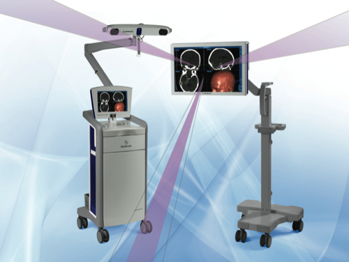
Stereotactic Craniotomy
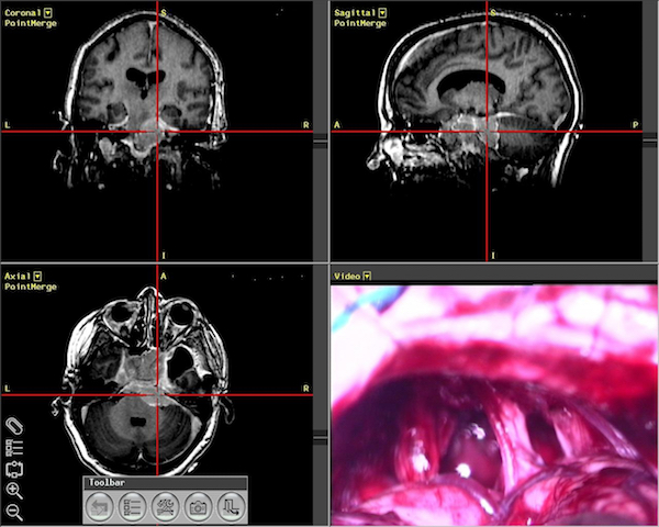
To localise a tumour within the brain, stereotaxis is often used. A preoperative MRI or CT scan is performed with fiducials (small green stickers) placed on the scalp. The scan data is uploaded to a special computer in the operating theatre, and a 3-Dimensional model of the head is created. Using a special system (similar in principle to your car's satellite navigation) the computer can assist the surgeon to localise a tumour accurately, thus improving overall efficiency, accuracy and outcomes.